- Share
- Share on Facebook
- Share on LinkedIn
Expertise
The imaging and flow cytometry platform develops methodologies for monitoring and quantifying mitochondrial structure and function, and parameters linked to energy metabolism of living cells:
- mitochondrial membrane potential
- opening of the permeability transition pore (NADH autofluorescence, unique know-how)
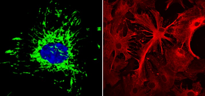
- organization of the mitochondrial network
- variations in ATP concentration
- oxidative stress
- calcium, protein/protein interactions (Duolink Method)
- visualization of various cellular interactions (cytoskeleton, endoplasmic reticulum)
- cell viability
The facility is part of the Grenoble network "Imagerie en Sciences du Vivant" and consists of:
- Confocal and fluorescence microscopy
- Cytometry analysis
- Analysis of microscopic images
Facilities
Quality approach according to ISO 9001 standard
Training and supervision of users, collaboration or service, acces to the material depending on the user skills (prices according to the applicable rates).
Material
2 confocal microscopes
Leica TCS SP2 AOBS: laser UV (351-364nm) + 3 visibles (488nm with AOTF (458-476-488-496-514nm), 543nm et 633 nm), DIC.
Leica TCS CSU SP8 : 3 lasers visibles (488, 552, 638nm) , Hybride detector.
1 cytometry analyzer
BD LSR Fortessa : 2 lasers (488nm and 532nm)
Image analysis station
Image J, Icy, Volocity
Contact
Cécile COTTET
Ingénieur INSERM
cecile.cottet univ-grenoble-alpes.fr (cecile[dot]cottet[at]univ-grenoble-alpes[dot]fr)
Phone number: +33 (0)4 76 63 54 80
- Share
- Share on Facebook
- Share on LinkedIn